Wednesday, December 13, 2023

Exciting New Results in the Development of an Anti-Malaria Vaccine


An exciting recent development has been reported in the prestigious journal Science regarding the development of a anti-malaria vaccine that has been shown to substantially reduce childhood victims of this disease. This vaccine is referred to as RTS, S. or Mosquirix and made by GSK.

Analysis of the efficacy of this vaccine approved to combat the death of the young children demonstrated a 13% drop in mortality during a nearly 4 year duration. This result was reported by the World Health Organization (WHO). In addition it was also found that there was a 22% reduction in the incidence of severe malaria in children young enough to receive a three-shot series.

According to John Tanko Bawa, director of the malaria vaccine implementation at the Program for Appropriate Technology in Health (PATH) stated, “The RTS,S malaria vaccine is already saving lives.” Furthermore he noted that, “What we have seen is a considerable impact of a vaccine described as having modest efficacy.”

The results of this analysis is so impressive that it has been estimated that the mortality decline could ultimately save tens of thousands of lives if RTS,S, is more broadly utilized.

In regard to the actual mode of action of RTS,S vaccine, it binds to the circumsporozoite protein on the surface of P. falciparum parasite before it infects liver cells disrupting its life cycle so that it is unable to infect circulating red bloods where it exerts its deadly effect – a pre-erythrocytic vaccine.

This is a profoundly important development in regard to the control of the spread of malaria among susceptible human populations.

Friday, March 3, 2023

Breast Cancer Overview - 2023


Introduction

It is currently estimated that one in eight women (12.5%) in the United States will be diagnosed with breast cancer.  For example, In the year 2014 232,670 new breast cancer cases and 40,000 deaths were reported for women living in the United Sates. Age is the strongest risk factor for breast cancer. Surprisingly, breast cancer begins to rise in the third decade of life.  This unusual aspect of breast cancer, is postulated to be related to the effects of ovarian hormones – especially estrogen and progesterone - on breast tissue. More than 2/3 of all new cases occur after the age of 55 and women older than 65 have a relative risk greater than 4.0 when compared with those younger than 65.

For this reason, it is imperative that causative agents responsible for the transformation of normal breast tissue cells to a cancerous state be more fully understood; that women be encouraged to undergo the appropriate screening and health checkups; and that more anti-breast cancer therapies be developed to combat this disease.

Additional Risk Factors

In addition to the endogenous ovarian hormones as cited earlier, there are the following factors that may play a significant role in the etiology of breast cancer –

  • Genetic factors such as the BRCA1 and BRCA2 genetic mutations and family history of the disease pointing to genetic factors that are poorly understood.
  • High levels of HER2 (HER2+) – an epidermal growth factor –can trigger uncontrolled cell division in breast tissue.  TCHP is a combination drug treatment that includes docetaxel, carboplatin, trastuzumab, and pertuzumab. These are drugs that people take intravenously to kill cancer cells if they have early-stage HER2+ breast cancer.
  • Reproductive history
  • High dose radiation to the chest
  • High dose hormone therapy
  • Obesity
  • Alcohol Consumption
  • Environmental factors related to the abundance of carcinogenic compounds that permeate the environment.

Endogenous Estrogen Levels and the Etiology of Breast Cancer

The Data accumulated in the past few decades indicate that endogenous estrogens play a very important role in regard to the etiology of breast cancer.  For this reason it is important to understand how estrogens are produced and metabolized in the body.  Estrogen and Progesterone are steroid hormones, and the first step involving steroidogenesis in the human ovary is the transport of their precursor, cholesterol into the mitochondria.  This is followed by a number of enzyme-mediated steps that lead to the formation of Pregnenolone that is the precursor for all steroid hormones, and eventually to estrogen.

In premenopausal women, estradiol synthesized in the ovaries is the most predominant form; whereas in postmenopausal women, estrone is the most prevalent and is synthesized in the peripheral tissue.  Estrone is reversibly converted to estradiol through an enzyme-mediated reaction.  Testosterone, in turn, is converted to estradiol by the action of aromatase enzyme in the peripheral tissues.  Aromatase is the enzyme that mediates the rate-limiting step in the conversion of androgens like testosterone into estrogens.  On account of the paramount importance of this metabolic step, pharmaceuticals that can effectively block aromatase activity have proven to be important aspect of the treatment of estrogen-dependent diseases such as breast cancer, endometriosis, and endometrial cancer.

It has been well established that active genes within the DNA serve as molecular blueprints for the production of unique proteins.  The steps in chemical metabolism within all the cells in the human body are mediated by specific enzymes that act as highly specialized chemical catalysts.  Enzymes are proteins.  Without enzymes life on earth would not be possible.  The dictum, “one gene one enzyme” can be applied universally throughout life.

It is important to keep in mind that by the very nature of their integration into DNA, genes are inheritable.  It is not uncommon to find polymorphisms within genes that are slight variations in the structure of those genes and what is referred to as single nucleotide polymorphisms (SNPs) that represent a singular change in the gene.  These variations in genetic structure produce corresponding variations in the proteins that are encoded in the genes that are the blueprints for these proteins.

Given this overall view, genetic research in regards to breast cancer is guided by an investigation of the genes that encode the structure of the enzymes involved in estrogen production.  The driving motivation of some of this work is to find the answer to the following question – Could the polymorphisms and SNPs in the genes responsible for the production of estrogens that are found in breast cancer patients result in an over-production of estrogens?  Secondly, could this over-production trigger the onset of breast cancer?

Clinical data that reinforces the primacy of estrogens in the onset of breast cancer are the following:

  • Bilateral oophorectomy (ovary removal) significantly reduces breast-cancer risk, and that risk reduction is greater if the ovaries are removed earlier in life.
  • In addition, some of the well-established risk factors for breast cancer, including early onset of menarche (menstruation) (<12 years), late menopause (>55 years), nulliparity or having child late in life, are related to lifetime exposure of breast tissue to sex hormones.
  • Approximately 2/3 of breast tumors are estrogen receptor (ER) positive (ER+) and responsive to circulating estrogens, and that almost all ER negative (ER−) cases are resistant to endocrine therapy, it is important to elucidate the specific mechanisms by which estrogens are related to elevated breast cancer risk.

In fact, circulating primary hormones in postmenopausal women, increased circulating concentrations of estradiol, estrone, estrone-sulfate, and androstendione have been shown to correlate with higher breast cancer risk. A thorough analysis of 663 women who developed breast cancer and had not received any hormonal-based therapy, demonstrated that the risk of breast cancer significantly increased with higher endogenous levels of total estradiol, free estradiol, estrone, estrone-sulfate, androstenedione, dehydroepiandrosterone (DHEA), dehydroepiandrosterone sulfate (DHEAS), and testosterone. Since this analysis was published, a few more prospective and case-control studies have been reported that have found similar results. It should be noted that the majority of populations studied were general populations with average breast cancer risk who were not taking any exogenous sex hormones.

In addition the levels of endogenous estrogens were studied in those patients that had a number of breast cancer risk factors including obesity, reproductive, demographic, and life style factors has been investigated by the Endogenous Hormones and Breast Cancer Collaborative Group in several studies. The results of these studies did not a show a statistical significance for the association between BMI (a metric whose value can be indicative of obesity) and breast cancer risk. However, in another cross-sectional analysis of 13 prospective studies by the same group, “estrogen and androgen levels were positively associated with obesity, smoking (15+ cigarettes daily) and alcohol consumption (20+g alcohol daily), and inversely linked with age.”

Although this summary does not include any data regarding the role of the level of endogenous androgens or progesterone in regard to the onset of breast cancer, the role of estrogens in the biology of breast cancer is very significant, and has led to the development of hormonal therapy medications as a way to limit the exposure of breast tissue to circulating estrogens. What follows is a more detailed look at these therapeutic approaches.

“Hormonal therapy medicines are used in four ways: (Jenni Sheng, MDJohns Hopkins University School of Medicine, Baltimore, MD)

“If the breast cancer is large and hormone receptor-positive, your doctor may recommend hormonal therapy before surgery to shrink the cancer. Treatments given before surgery are called neoadjuvant treatments, so hormonal therapy given this way is called neoadjuvant hormonal therapy.


“To reduce recurrence risk: If you’ve been diagnosed with early-stage hormone receptor-positive breast cancer, your treatment plan will include hormonal therapy after surgery and possibly other treatments to reduce the risk of the cancer coming back (recurrence). Treatments given after surgery are called adjuvant treatments, so hormonal therapy given this way is called adjuvant hormonal therapy.

“To stop advanced-stage cancer from growing: If you’ve been diagnosed with advanced-stage, hormone receptor-positive breast cancer, hormonal therapy can be used to help stop the cancer from growing.

“To reduce the risk of a first diagnosis: Hormonal therapy also can be used to reduce breast cancer risk in certain women who haven’t been diagnosed. Women with a much higher than average risk of breast cancer may take a hormonal therapy medicine preventively to reduce the risk of hormone receptor-positive breast cancer developing.

“How does hormonal therapy treat breast cancer?

Hormonal therapy medicines work in two ways:

  • by blocking estrogen production in the body


  • by blocking the effects of estrogen on breast cancer cells

“Hormonal therapy is not a treatment option for hormone receptor-negative breast cancer.

“It's important to know that hormonal therapy for breast cancer is different than hormone replacement therapy (HRT) for treating symptoms of menopause. HRT isn't used to treat breast cancer. HRT is taken by some women to treat troublesome menopausal side effects such as hot flashes and mood swings. HRT is used to raise estrogen levels that drop after menopause. HRT contains estrogen and can contain progesterone and other hormones. Hormonal therapy for breast cancer is exactly the opposite — it blocks or lowers estrogen levels in the body.

“Types of hormonal therapy to treat breast cancer

“There are three main types of hormonal therapy medicines used to treat breast cancer:

                     Aromatase inhibitors stop the body from making estrogen.


                     Selective estrogen receptor modulators (SERMs) block the action of estrogen on certain cells.


                     Selective estrogen receptor downregulators (ERDs) block the action of estrogen on certain cells.


                     Aromatase inhibitors

“Aromatase inhibitors lower estrogen levels by stopping the enzyme aromatase from changing other hormones into estrogen. In estrogen receptor-positive breast cancer, the hormone estrogen can stimulate the growth of breast cancer cells.


“There are three aromatase inhibitors used to treat breast cancer:


                     Arimidex (chemical name: anastrozole)


                     Aromasin (chemical name: exemestane)


                     Femara (chemical name: letrozole)


“Selective estrogen receptor modulators (SERMs)

“Selective estrogen receptor modulators (SERMs) block the effects of estrogen on breast cancer cells by sitting in the estrogen receptors. If a SERM is in the estrogen receptor, estrogen can’t attach to the cancer cell and the cell doesn’t receive estrogen’s signals to grow and multiply.

“There are three SERMs used to treat breast cancer:


                     Tamoxifen in pill form, also called tamoxifen citrate (brand name Nolvadex), and in liquid form (brand name: Soltamox)


                     Evista (chemical name: raloxifene)


                     Fareston (chemical name: toremifene)


“Selective estrogen receptor downregulators (SERDs)

“Selective estrogen receptor downregulators (SERDs), much like SERMs, block the effects of estrogen on breast cancer cells by sitting in the estrogen receptors. SERDs also lower the number of estrogen receptors and change the shape of breast cell estrogen receptors so they don’t work as well. There are two SERDs used to treat breast cancer:


                     Faslodex (chemical name: fulvestrant)


                     Orserdu (chemical name: elacestrant)


“Hormonal therapy side effects

Each hormonal therapy medicine may cause different side effects.

The most common side effects of the aromatase inhibitors are:

                     joint and bone pain


                     hot flashes


                     fatigue


                     weakness

“The most common side effects of the SERMs are:

                     hot flashes


                     vaginal discharge


                     mood swings


                     fatigue


“The most common side effects of the SERDs are:

                     nausea


                     bone pain


                     fatigue


                     hot flashes


                     injection site pain (for Faslodex only)


“For many years, women took hormonal therapy for five years after surgery for early-stage, hormone receptor-positive breast cancer. In most cases, the standard of care is five years of tamoxifen, or two to three years of tamoxifen followed by two to three years of an aromatase inhibitor, depending on menopausal status.

“Recent research has found that in certain cases, taking tamoxifen for 10 years instead of five years after surgery lowered a woman’s risk of recurrence and improved survival.

“In most cases, a post-menopausal woman diagnosed with early-stage, hormone receptor-positive breast cancer would take an aromatase inhibitor for five years after surgery to reduce the risk of recurrence. After that, if breast cancer had been found in the lymph nodes, called node-positive disease, a woman would take an aromatase inhibitor for an additional five years, for a total of 10 years of hormonal therapy treatment.

“Doctors call taking hormonal therapy for 10 years after surgery extended adjuvant hormonal therapy.

“Ovarian suppression or removal

“In pre-menopausal women, most of the estrogen in the body is made by the ovaries. In some cases, medicine may be used to stop the ovaries from functioning temporarily, called ovarian suppression or ovarian shutdown. Two medicines commonly used are:

                     Zoladex (chemical name: goserelin)


                     Lupron (chemical name: leuprolide)

 “These medicines are given as injections once a month for several months or every few months. They can be used alone or in combination with other hormonal therapy medicines to treat pre-menopausal women.

“Once you stop receiving the medicine, your ovaries usually begin functioning again. The time it takes for the ovaries to recover varies from woman to woman.

“Some women with a much higher than average risk of breast cancer may choose to have their ovaries removed, called prophylactic or preventive ovary removal, either before or after being diagnosed with breast cancer.”

This article is designed to summarize the known relationship between estrogen levels and the majority of breast cancers (ER+).  It is important to keep in mind that this area of scientific, medical and clinically-based research is constantly generating new data, and the findings presented above do not represent the last word on the understanding of this devastating illness.


Thursday, January 19, 2023

An approach to Cancer Treatment Combining Immunotherapy and Chemotherapy


Immunotherapy is an approach that focuses on and improving the body’s inherent capacity to attack tissue cells that have been transformed into cancerous cells. However, the current drawback of this approach is its inability to effectively distinguish the target cell from neighboring healthy cells. This is especially true for cancers that are derived from oncogene expression. Chemotherapy that has long been the mainstay of cancer treatment has a serious limitation of its own – actively growing cancer cells often develop a resistance to this kind of drug treatment.
Takmitsu Hattori and his colleagues at the Laura and Issac Perlmutter Cancer Center at New York University have attempted to overcome this impasse by developing a methodology that combines these two methodologies. Their approach is to effectively create a neoantigen, a hapten-peptide conjugate, which can preferentially combine with cancer cells and serve as a target for selective elimination.

These research scientists treated lung cancer with sotorasib – a drug that specifically targets KRAS – an oncogene that is involved in cell signaling pathways that are in involved in the control of cellular proliferation, cell development and cell death. Subsequently, they administered synthetically created antibodies designed to recognize the neoantigens that were the product of KRAS generated protein that had been bound to sotorasib. These administered antibodies were shown to have selectively killed even the sotorasib-resistant cells while leaving normal cells intact.


Sturcture of Sotorasib

These results show a great deal of promise in regard to adding to the repertoire of cancer treatment modalities.


Tuesday, January 17, 2023

A Vaccine for Respiratory Syncytial Virus (RSV) Virus May Soon be Available

This fall the general population has been plagued by an increased incidence of illnesses caused by three distinct viruses - COVID 19 (Coronavirus), Respiratory Syncytial Virus (RSV) and the Influenza Virus.

Infants and the elderly can be particularly impacted by severe illness with RSV. RSV is a negative-strand RNA virus. The term syncytial comes from the fact that host cells infected by the virus fuse and form large cells, syncytia. Although RSV infection is quite common and the respiratory issues it produces are usually not severe and of short durations, for infants and the elderly, it can produce severe and dangerous symptoms that may include difficulty breathing, abnormal respiratory sounds and coughing and wheezing that does not stop.


This fall, hospitalization of infants and the elderly with RSV has been particularly devastating. This reality has accelerated the development of a safe and effective vaccine against this virus. It has recently be reported in the prestigious journal, Science that two large trials have proven the efficacy of two vaccines against RSV infection. The data demonstrate that either vaccine can protect both infants and individuals over the age of 60. One of these vaccines also was found to protect infants for up to six months when given to women in the latter stage of pregnancy who could pass the antibodies induced by the vaccine to the fetus.

Some fifty years ago an initial attempt to develop a vaccine against RSV proved unsuccessful; a chemically inactivated intact virus was used in this particular vaccine. Not only did the vaccine fail to elicit a robust response to RSV infection, it also led to some fatalities and even worsened the symptoms of those treated from subsequent RSV infection.

It was reported that, “The new vaccines avoid this problem by relying on a key advance made by Barney Graham and co-workers at the National Institute of Allergy and Infectious Diseases in 2013”

They discovered that a key viral protein located on the surface of the viral membrane interacts with a particular receptor on the cell membrane of the target tissue and changes its shape allowing it to gain entry into the host cell. The team working on vaccine development led by Doctor Graham currently at the Morehouse School of Medicine (Atlanta, Georgia) used this knowledge to modify the viral protein so it remains fixed in this modified state. Once this modified antigen was introduced into the vaccine, it resulted in the production of higher levels of effective antibodies. This good news was established as a result of clinical trials run by GSK and Pfizer. This result illustrates how the knowledge gained from the study of viral biology can prove efficacious in the development of treatments against infection.


Friday, November 11, 2022

The Role of Microbial Metabolites in the Etiology of Colon Cancer

 It has been shown that a number cancers are caused by pathogens as the table below demonstrates


Disease

Causative Agent


T-cell Leukemia

HTLV-1 Human Retrovirus

Cervical Cancer

Human Papilloma Virus (HPV) 

Liver Cancer

Hepatitis B and Hepatitis C Virus (HBV and HCV)

Stomach Cancer

H.pylori – a Bacteria


This growing evidence involving the role of pathogens in the etiology of various cancers has led to numerous studies that look at a possible role of enteric microorganisms that naturally inhabit the human gut in the etiology of colon cancer.  The result of this kind of investigation has revealed that so-called genotoxic gut bacteria, whose metabolic product(s) can damage or mutate human DNA, may well be drivers of colorectal cancer (CRC) parthenogenesis.

Moreover, it has been recently reported in the prestigious journal, Science, by Dr. Cao and his colleagues that patients presenting with inflammatory bowel disease (IBD) – a condition that can lead to CRC – show the evidence of  diverse populations of  bacterial strains that can exert genotoxic activity.

 In this study, a particular bacterium, Monganella morganii, proved pf particular interest.  This organism is a Gramnegative bacterium that was discovered as an enriched population among individuals suffering from IBD and CRC.  The organisms of this type was identified as belonging to a class of bacteria that produce indolimines.  This is of special importance since indolimines have been found to promote tumor growth in mice.

 In addition to these findings, “Among the most well-described genotoxic bacteria are enterotoxigenic Bacteroides fragilis (ETBF) (3) that produce Bacteroides fragilis toxin (BFT), strains of Escherichia coli or other bacteria that produce colibactin (4), and Campylobacter jejuni strains that express cytolethal distending toxin (CDT) (5). The DNA-damaging properties of these strains and their toxins vary considerably, ranging from alkylating DNA interstrand cross-links for colibactin to deoxyribonuclease (DNase) activity for CDT (1, 4, 5). Intriguingly, ETBF and E. coli producing colibactin have been linked to IBD and CRC development using in vitro and in vivo models.”

The contribution of Dr. Chao and his colleagues made as cited in their study was the successful elucidation and characterization of the existence of some 18 strains of strains of enteric bacteria that actively produce genotoxins out of the 122 strains that were isolated for examination.

In the final analysis, the correlation between genotoxins found in the gut and the onset of IBD and CRC is of immense value and points to an avenue for future studies that may be of profound importance in regard to the ultimate prevention and treatment of these diseases.


Saturday, October 1, 2022

Immunotherapeutic Methodology Designed to Treat Lupus

Lupus is a disease in which the patient manifests certain symptoms that are a result of an autoimmune disease in which the patient’s immune system is producing antibodies against the individual’s own tissue(s).

The following is an overview of this condition as described on the website of the Mayo Clinic – (https://www.mayoclinic.org/diseases-conditions/lupus/symptoms-causes/syc-20365789)

“Lupus is a disease that occurs when your body's immune system attacks your own tissues and organs (autoimmune disease). Inflammation caused by lupus can affect many different body systems — including your joints, skin, kidneys, blood cells, brain, heart and lungs.

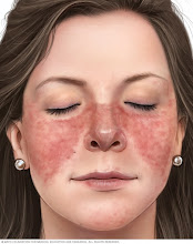
“Lupus can be difficult to diagnose because its signs and symptoms often mimic those of other ailments. The most distinctive sign of lupus — a facial rash that resembles the wings of a butterfly unfolding across both cheeks — occurs in many but not all cases of lupus.

“Some people are born with a tendency toward developing lupus, which may be triggered by infections, certain drugs or even sunlight. While there's no cure for lupus, treatments can help control symptoms.

Symptoms

Red, butterfly-shaped rash on nose and cheeks


 No two cases of lupus are exactly alike. Signs and symptoms may come on suddenly or develop slowly, may be mild or severe, and may be temporary or permanent. Most people with lupus have mild disease characterized by episodes — called flares — when signs and symptoms get worse for a while, then improve or even disappear completely for a time.

The signs and symptoms of lupus that you experience will depend on which body systems are affected by the disease. The most common signs and symptoms include:

Fatigue

Fever

Joint pain, stiffness and swelling

Butterfly-shaped rash on the face that covers the cheeks and bridge of the nose or rashes elsewhere on the body

Skin lesions that appear or worsen with sun exposure

Fingers and toes that turn white or blue when exposed to cold or during stressful periods

Shortness of breath

Chest pain

Dry eyes

Headaches, confusion and memory loss”


Autoimmune diseases as a class of ailments have been historically exceedingly difficult to treat. However, impressive inroads have been made in recent years using the rapidly advancing techniques embodies in immunotherapy. It has been reported in a recent issue of the prestigious scientific publication, Nature Medicine, from a medical team in Germany that five patients, four women and a man, have been successfully treated with their own immune cells that have been genetically engineered.

This technique involves isolating the patient’s own T cells – a subset of the immune system’s cellular repertoire – and genetically modifying them so that they would recognize those B cells that are involved in the autoimmune response unique to lupus and attack them by binding to a specific cell surface protein (antigen). This kind approach is referred to as T cell (CAR-T) therapy.

In this case, all five patients tolerated the therapy and their lupus-caused impaired organ function either improved or was resolved. As a result, these patients were no longer required to take immune-suppressive medication.

These results are quite encouraging and certainly are promising in regard to the treatment of lupus.

Friday, May 27, 2022

The Beginning of Cellular Life on Planet Earth - An Hypothesis

A Possible Structure for a Primitive Cell- Biology Libre Texts

A necessary first step in examining the evolution of cellular-based life from the pre-biotic world is to discern what constitutes the makeup of cells in the broadest possible terms. In deconstructing cell structure from the viewpoint of prokaryotes, I propose the following necessary prerequisites for a living cell:

· Cell Membrane to delineate the cell and protect it from the local environment

· Cell infrastructure composed of the most basic structural components including actin, myosin, etc.

· Source of readily available energy

· Proteins especially enzymes involved in catabolic and anabolic activities

· Signal transduction pathways that allow for both intracellular and extra-cellular communication.

· Nucleic Acids: DNA and RNA to serve as information stores for the cell.

· Mechanisms for DNA and cellular replication.

Any attempt to propose a mechanism by which primordial cell-like structures evolved into the complex cells that exist today, strongly suggests a gradual stepwise process that took eons to accomplish. It also suggests, in my mind, that the process would involve steps in which cellular organization would grow in complexity from the level of simple molecules (substrates) through proteins and nucleic acids and finally through protein-nucleic acid interaction to the encoding of the genetic material. In this way, selection pressures and processes would enhance each succeeding step.

Taking all this into account, I propose the following model for the evolution of cellular life from primordial beginnings.

Elements of the Hypothesis:

There existed an aqueous environment (possibly shallow ponds or along coastal regions or possibly the sea floor) where there was an abundance of nucleotides, fatty acids, amino acids, peptides and polypeptides. It is possible that some of this organic material may have been seeded by meteorites.
In these organic-enriched regions, conditions were appropriate for the spontaneous formation of cell-like structures.
 
These cell-like structures developed semi-permeable membranes formed from the spontaneous assembly of proteins and lipids (probably a more primitive structure than found in present day cells) and highly permeable to dissolved organic matter in the local environment.
 
The local environment was such that amino acids, nucleotides, fatty acids and carbohydrates could readily penetrate the cell membranes of these primordial cells and concentrate there.
 
Ambient conditions including oxygen concentration, temperature, abundance of ammonia and methane made the spontaneous synthesis of proteins and nucleic acids not only possible but highly likely.
Assuming that spontaneous formation of tRNAs were a likely scenario, these tRNAs could bind to their appropriate amino acids. These amino acid carriers collided with each other and resulted in the formation of random polypeptide chains. Subsequently, polypeptides that were capable of binding to carbon sources such as glucose stabilized these small proteins and gave them a competitive advantage over more non-specific proteins. Since the metabolic pathway for glucose metabolism is universal to all life, one must assume that glucose was abundant in pre-biotic times. This same argument can be applied to the presence of ADP/ATP, since this molecule is the essential ingredient for all energy sustaining life activities.
 
Some of these selected proteins also possessed catalytic capabilities and were able to breakdown carbon rich substrates and ultimately capture energy in ATP molecules. This energy may have been used in accelerating the synthesis of more complex molecules and intra-cellular structures, the precursors of cellular organelles.  
 
There is mounting evidence that strongly suggests that RNA may have played a pivotal role in information storage in the early evolution of cellular life. As I have postulated above, tRNAs may have been abundant. Additionally, evidence for the role of RNA in information storage includes:
 
  • The discovery of RNA that possesses catalytic activity referred to as ribozymes. There is a ribozyme that has been found in the core of ribosomes.   
  • The discovery of small pieces of RNA that can readily bind to a variety of organic molecules and that are found on the ends of mRNA in prokaryotes. These pieces function as switches that can turn translation on or off and are referred to as riboswitches. 
  • Double-stranded RNA that can silence gene transcription in a complex referred to as RISC.
  • What is now referred to as the anti-codon region of tRNA may have been used to make mRNA possibly happening spontaneously utilizing an environment rich in small pieces of RNA or assisted by a ribozyme. These nascent mRNAs served as templates for the further synthesis of specific and biologically valuable proteins. Whether or not such associations are possible today in conditions that simulate the pre-biotic environment would need to be tested. It is possible that “ancient” RNA had a different structure than the current form. This early mechanism was probably inefficient and prone to error.
 
These early cells were infiltrated by a competing entity that gradually assumed a symbiotic relationship and was to become what is now referred to as ribosomes. These structures contributed a much more efficient mechanism for the synthesis of proteins.  In addition, the mitochondria found in eukaryotic cells and the chloroplasts found uniquely in plant cells have their own nucleic acid and most probably were once independent organisms that also assumed a symbiotic relationship with their host cells.
 
Messenger RNAs were no longer able to sustain the growing complexity of cell life as embodied in metabolism and energy transfer mechanisms. A more highly conserved store of information was required. The appearance of an enzyme capable of using mRNA as a template to make highly stable double-stranded DNA encouraged the further development of cellular complexity and evolution. This transition was necessitated by the fact that the extra-cellular environment was no longer as rich in nutrients and building materials as was previously the case. It has become clear that large portions of the genome of humans and other complex organisms are made up of retrotransposons. There are relatively small pieces of DNA that code for reverse transcriptases that allow for copying of these segments and ultimately inserting them in other places in the genome. These were originally discovered by McClintock and referred to as so-called “jumping genes.” Integration of these pieces in the promoter or structural regions of active genes can have profound impacts on gene expression. Certain diseases have been associated with this process. Furthermore, there are retrotransposons that have been conserved among and between organisms. This suggests that the increasing complexity of the genome as seen in evolution may be in large due to retrotransposons. In addition, retrotransposons have many characteristics similar to retroviruses suggesting that retroviruses may have played a significant role in delivering novel genetic material to the genome.
In conclusion, the particular scenario I have outlined represents one possible pathway that may have taken place that was responsible for the evolution of cellular life as we know it from prebiotic conditions that existed on planet Earth billions of years ago.